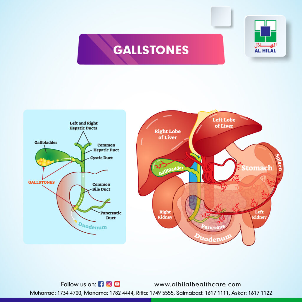
Gallstones are often made of cholesterol or bilirubin and can develop in the gallbladder or bile ducts. These stones can cause pain and other complications.
Stones Made Of Cholesterol: This is the most common type. Cholesterol gallstones are not related to cholesterol levels in the blood. Usually, your bile contains enough chemicals to dissolve the cholesterol excreted by your liver. But if your liver passes more cholesterol than your bile can dissolve, the excess cholesterol may form into crystals and, eventually, stones.
Stones Made Of Bilirubin: These are called pigment stones. They occur when red blood cells are destroyed, and too much bilirubin is in the bile. Certain conditions cause your liver to make a lot of bilirubin, including biliary tract infections, liver cirrhosis, and certain blood disorders. In addition, the excess bilirubin contributes to gallstone formation.
CAUSES OF GALL STONE:
The cause of gallstones varies:
- Obesity
- Females are more common
- Age more than 40
- In pregnancy, progesterone decreases the contractility of the gallbladder leading to stas
- Rapid weight loss from eating a very low-calorie diet or after weight loss surgery
- Receiving nutrition through a vein for an extended period (intravenous feedings)
- Taking birth control pills
- Diabetes
- Bone marrow or solid organ transplant
SYMPTOMS OF GALL STONES:
Gallstones may cause no symptoms or signs. If a gallstone lodges in a duct and causes a blockage, the resulting symptoms and signs may include:
- Rapidly intensifying and sudden pain in the upper right portion of the abdomen
- Back pain between your shoulder blades
- Pain in your right shoulder
- Nausea or vomiting
- Other digestive problems include indigestion and heartburn
- Fever and chill
- Yellow skin or eyes
- Dark urine and light-colored stool
GALLSTONE DIAGNOSIS:
Blood Investigation: Blood tests may reveal infection, jaundice, pancreatitis, or other complications caused by gallstones.
Complete blood count (CBC), Liver Function Test, Serum Amylase & Lipase.
Ultrasound: It is the gold standard to look for gallstones because it is simple and non-invasive. Ultrasound is very good at highlighting gallstones within the gallbladder, as well as features, such as a thickened gallbladder wall, that point to inflammation of the gallbladder (acute cholecystitis)
Magnetic Resonance Cholangiopancreatography (MRCP): MRCP test uses a magnetic field and pulses of radio wave energy to take pictures of the inside of your body, including the liver and gallbladder.
Cholescintigraphy (HIDA scan) helps in the diagnosis of several diseases and conditions, such as:
- Gallbladder Inflammation (Cholecystitis)
- Bile Duct Obstruction
- Congenital abnormalities in the bile ducts, such as biliary atresia
- Postoperative complications, such as bile leaks and fistulas
- Assessment of liver transplant
Endoscopic Retrograde Cholangiopancreatography (ERCP): In this method, your doctor runs an endoscope through your mouth to your small intestine. They will inject a dye so they can see your bile ducts on a camera in the endoscope. They can usually take out any gallstones that have moved into the ducts.
Endoscopic Ultrasound: This test combines ultrasound and endoscopy to look for gallstones.
COMPLICATIONS OF GALLSTONES:
Gallstones can cause serious problems, including:
Gallbladder Inflammation (Acute Cholecystitis): This happens when a stone blocks the gallbladder so it can’t empty. It causes constant pain and fever.
It blocked bile ducts. This can cause fever, chills, and yellowing of skin and eyes (jaundice). In addition, if a stone blocks the duct to the pancreas, that organ may become inflamed (pancreatitis).
Infected Bile Ducts (Acute Cholangitis): A blocked duct is more likely to get infected. The bacteria spread to the bloodstream can cause a dangerous condition called sepsis.
Gallbladder Cancer: It’s rare, but gallstones raise your risk of this kind of cancer.
TREATMENT FOR GALL STONES:
Most of the time, there is no need for treatment for gallstones unless they cause pain. Sometimes it can pass gallstones without even noticing.
Acute Cholecystitis: Treatment for cholecystitis usually involves a stay in the hospital to control the inflammation. Sometimes, surgery is needed.
The initial treatment is admission, fluid management, analgesic, and IV antibiotics.
Usually, surgery is indicated after four to six weeks of medical treatment.
Laparoscopic cholecystectomy is a minimally invasive procedure because it uses several small incisions instead of one large one. A laparoscope is a narrow tube with a camera. This surgical tool is inserted through one incision. The camera allows seeing the gallbladder on a TV screen. The gallbladder is then removed through another small incision.
ADVANTAGES OF LAPAROSCOPIC CHOLECYSTECTOMY:
- Less pain
- Lower risk of complications
- Quicker recovery and return to regular activities – daycare procedure
- Smaller wounds and scar
Open Cholecystectomy is typically performed when the gallbladder is inflamed, infected, or scarred. However, this surgery may also happen if complications occur during laparoscopic cholecystectomy.
The Endoscopic Retrograde Cholangiopancreatography (ERCP) procedure is another minimally invasive option for removing gallstones.
Percutaneous Gallbladder Drainage involves placing a sterile needle into the gallbladder to draw out bile. A tube is then inserted to assist with additional drainage. This procedure isn’t typically the first line of defense and tends to be an option for individuals who may not be suited for other methods.
A COMPLICATION OF GALL BLADDER SURGERY:
Removal of the gallbladder (cholecystectomy) is considered a relatively safe procedure, but like all operations, there’s a small risk of complications
- Intra operation
- Bile leak
- Bleeding
- Injury to nearby structures, such as the liver, liver, and small intestine
- Post-surgery – infection (rare)
Oral dissolution therapy typically includes using the medications ursodeoxycholic acid and Chenodeoxycholic acid to break up gallstones. These medications contain bile acids, which work to break up the stones. This treatment is best suited for breaking up cholesterol stones.
CONCLUSION:
Cholelithiasis or gallstones are hardened deposits of digestive fluid that can form in the gallbladder. The gallbladder holds a digestive fluid known as bile that is released into the small intestine. Women are more affected, most of which are asymptomatic in patients with asymptomatic gallstones discovered incidentally, the likelihood of developing symptoms or complications. Asymptomatic gallbladder stones found in a normal gallbladder and standard biliary tree do not need treatment unless they develop symptoms. However, approximately 20% of these asymptomatic gallstones will develop symptoms over the years of follow-up. These gallstones may go on further to develop complications such as cholecystitis, cholangitis, choledocholithiasis, gallstone pancreatitis, and rarely cholangiocarcinoma. This activity reviews the etiology, presentation, evaluation, and management of cholelithiasis and reviews the role of the interprofessional team in evaluating, diagnosing, and managing the condition.